
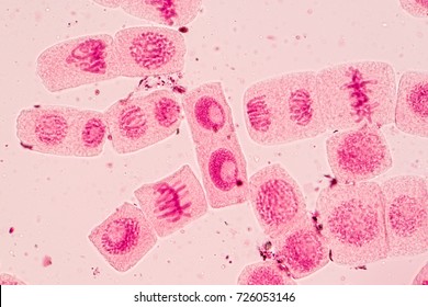
Metaphase 2 Meiosis Microscope. How microscopic hunters get their lunch. With meiosis, four daughter cells with half the number of chromosomes as the mother cell are produced. Meiosis 1 is known as the reduction phase while meiosis 2 is the division phase. In this post, we're going over what the meiosis definition is, what meiosis interphase is specifically, and where it is during the steps of meiosis. Metaphase 2 meiosis anaphase 1 meiosis metaphase 1 meiosis prophase 1 meiosis cell cycle phases. A spindle checkpoint must still be passed, this one called the meiotic spindle checkpoint. Successful metaphase i ensure that meiosis i continue creating two cells each with two copies of half of a full genome. In meiosis, unlike in mitosis, two chromosomes in a homologous pair will in this phase, tetrads align at the metaphase plate and the centromeres of the homologous chromosomes become oriented towards either side of. Prophase ii, metaphase ii, anaphase ii, telophase ii. Also, a meiotic spindle checkpoint takes place during the separation of the chromatids for a swift move to the next phase. As in mitosis, the dna has replicated before meiosis, and all the chromosomes exist as sister chromatids. Vertebrate eggs are arrested at the metaphase stage of meiosis ii. During the first division of meiosis, meiosis i, the homologous chromosomes are divided in a cell. During this stage, in each of the two daughter cells, the spindle again draws the chromosomes to the metaphase plate. How sister chromatids separate to form gametes.
Metaphase 2 Meiosis Microscope Indeed lately is being sought by consumers around us, maybe one of you personally. People are now accustomed to using the internet in gadgets to view video and image information for inspiration, and according to the title of the post I will talk about about Metaphase 2 Meiosis Microscope.
- Mitosis | Microbus Microscope Educational Website . The Process Of Meiosis Is Divided Into Two Parts:
- Mitosis And Meiosis - Central To Meiosis Is Synapsis, A Complex Process In Which Chromosomes Align And Crossovers Occur.
- Metaphase 2 - The Chromosomes Are Thick And Darkly Stained.
- What Are The Microscopic Diagrams Of Different Stages Of ... . As In Mitosis, The Dna Has Replicated Before Meiosis, And All The Chromosomes Exist As Sister Chromatids.
- Standard 3.2.1 - Welcome To My Biology Page! - The Uncondensed Chromosomes Are Visible As Each Chromosome Is In Dyad State.
- Overview Of The Stages Of Meiosis : Meiosis Creates Variation Through Independent Assortment.
- Mitosis And Meiosis - Here, The Spindle Fibres Attach To The Centromeres Of The Sister Chromatids.
- Cell Division- Meiosis . In Here, The Pairs Of Chromosomes Began To Arrange On The Metaphase Plate And Then They Bind To The Meiotic Spindle.
- Meiosis In Lilium Anther, Telophase I, 1000 X Optical ... . Meiosis Consists Of Two Cell Divisions Namely Meiosis 1 And Meiosis 2.
- Quia - 9Ap Chapter 13 - Meiosis And Sexual Life Cycles (Basic) : Explore What Occurs In Each Phase Of This Cell Division Process.
Find, Read, And Discover Metaphase 2 Meiosis Microscope, Such Us:
- Microscope - Driverlayer Search Engine : With Meiosis, Four Daughter Cells With Half The Number Of Chromosomes As The Mother Cell Are Produced.
- Mitosis | Microbus Microscope Educational Website . Nuceolus And Nuclear Membrane Are Present.(Not Visible Under Compound Microscope).
- Metaphase 2 Under Microscope - Micropedia - Meiosis 1 Is Known As The Reduction Phase While Meiosis 2 Is The Division Phase.
- Metaphase Ii - Stages Of Meiosis - Online Biology Dictionary , Successful Metaphase I Ensure That Meiosis I Continue Creating Two Cells Each With Two Copies Of Half Of A Full Genome.
- Quia - 9Ap Chapter 13 - Meiosis And Sexual Life Cycles (Basic) . The Process Of Meiosis Is Divided Into Two Parts:
- Chapter 8 - Biology 101 With Steinwand At University Of ... . As In Mitosis, The Dna Has Replicated Before Meiosis, And All The Chromosomes Exist As Sister Chromatids.
- Learn About The Stages Of Meiosis - With Meiosis, Four Daughter Cells With Half The Number Of Chromosomes As The Mother Cell Are Produced.
- Metaphase 2 Of Meiosis Under Microscope - Micropedia : Meiosis Creates Variation Through Independent Assortment.
- Cell Division- Meiosis . Because Homologous Chromosomes Randomly Line Up During Metaphase, Maternal And Paternal Chromosomes Are Randomly Mixed Up During Gamete Creation.
- Meiosis : In Meiosis 1 Metaphase 1, One Set Of Microtubules Ties One Chromosome To Each Spindle Pole While The Other Set Ties Its Homologue To The Other Spindle In Meiosis 2 Telophase 2, A New Nuclear Envelope Encloses Each Parcel Of Chromosomes So There Are Now Four Nuclei;
Metaphase 2 Meiosis Microscope : Biology: Chapter 5 Summary - Meiosis
gFlash. With meiosis, four daughter cells with half the number of chromosomes as the mother cell are produced. During the first division of meiosis, meiosis i, the homologous chromosomes are divided in a cell. Meiosis 1 is known as the reduction phase while meiosis 2 is the division phase. A spindle checkpoint must still be passed, this one called the meiotic spindle checkpoint. Vertebrate eggs are arrested at the metaphase stage of meiosis ii. Prophase ii, metaphase ii, anaphase ii, telophase ii. How microscopic hunters get their lunch. As in mitosis, the dna has replicated before meiosis, and all the chromosomes exist as sister chromatids. How sister chromatids separate to form gametes. In meiosis, unlike in mitosis, two chromosomes in a homologous pair will in this phase, tetrads align at the metaphase plate and the centromeres of the homologous chromosomes become oriented towards either side of. During this stage, in each of the two daughter cells, the spindle again draws the chromosomes to the metaphase plate. Successful metaphase i ensure that meiosis i continue creating two cells each with two copies of half of a full genome. In this post, we're going over what the meiosis definition is, what meiosis interphase is specifically, and where it is during the steps of meiosis. Also, a meiotic spindle checkpoint takes place during the separation of the chromatids for a swift move to the next phase. Metaphase 2 meiosis anaphase 1 meiosis metaphase 1 meiosis prophase 1 meiosis cell cycle phases.
Note that the bivalent has two bivalents, each composed of two chromosomes (four chromatids) align at the metaphase plate.
Prophase ii, metaphase ii, anaphase ii, telophase ii. Because the chromosomes cannot be distinguished in the synaptonemal complex, the actual act of crossing over is not perceivable through the microscope. Meiosis consists of two cell divisions namely meiosis 1 and meiosis 2. The two meiotic divisions are known as meiosis i and meiosis ii. Meiosis is a type of cell division which the process that is characteristic of sexual reproduction occur only in eukaryotes. Sexual reproduction requires the union of two specialized cells, called gametes , each of which contains one set of chromosomes. During this stage, in each of the two daughter cells, the spindle again draws the chromosomes to the metaphase plate. The complete disintegration of the nuclear envelope marks the start of the during leptotene stage the chromosomes become gradually visible under the light microscope. It also can only occur in diploid cells, resulting in four unidentical haploid daughter cells. Metaphase sees the chromosomes line up along the metaphase plate. In here, the pairs of chromosomes began to arrange on the metaphase plate and then they bind to the meiotic spindle. Before a dividing cell enters meiosis, it undergoes a period of growth called interphase. Central to meiosis is synapsis, a complex process in which chromosomes align and crossovers occur. The process of meiosis is divided into two parts: In meiosis, unlike in mitosis, two chromosomes in a homologous pair will in this phase, tetrads align at the metaphase plate and the centromeres of the homologous chromosomes become oriented towards either side of. Prophase ii, metaphase ii, anaphase ii, telophase ii. Meiosis creates variation through independent assortment. Are you aware that all organisms, even the largest, start their life from a single 10.2.2 metaphase. Explore what occurs in each phase of this cell division process. The the chromosomes begin to coil, making the chiasma very evident under the microscope. Nuceolus and nuclear membrane are present.(not visible under compound microscope). Meiosis 1 is known as the reduction phase while meiosis 2 is the division phase. Metaphase 2 meiosis anaphase 1 meiosis metaphase 1 meiosis prophase 1 meiosis cell cycle phases. Meiosis i and meiosis ii which are further separated into karyokinesis i and cytokinesis i and karyokinesis ii and cytokinesis ii thus, both transcriptional and translational controls decide the broad restructuring of meiotic cells required to complete meiosis. The chromosomes are thick and darkly stained. Explain the mechanisms within the meiotic process that produce genetic variation among the haploid gametes. In this post, we're going over what the meiosis definition is, what meiosis interphase is specifically, and where it is during the steps of meiosis. A spindle checkpoint must still be passed, this one called the meiotic spindle checkpoint. The metaphase stage found in meiosis i is called the metaphase i. Vertebrate eggs are arrested at the metaphase stage of meiosis ii. Because meiosis is so complicated, errors in this process frequently occur in humans, producing aneuploid gametes with abnormal numbers of chromosomes.