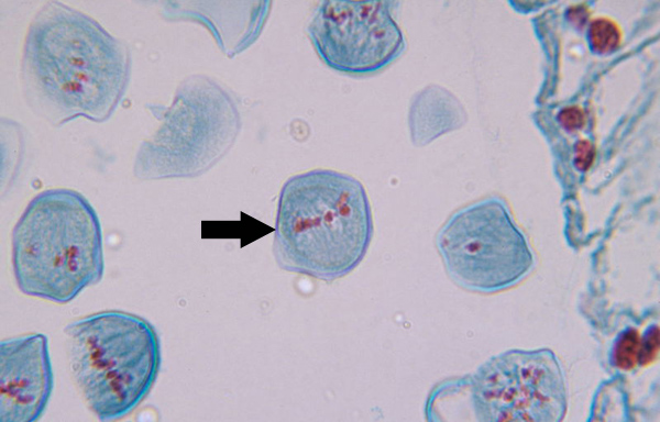
Meiosis Stages Seen Under Microscope. Anaphase usually only lasts a few moments and appears dramatic. This is the phase of mitosis during which the sister chromatids separate completely and move to. This video takes you through microscope images of cells going through mitosis and identifies the different phases under the microscope and on a micrograph. Two successive divisions without any dna replication. Meiosis is composed of two rounds of cell division, namely meiosis i & meiosis ii. At the beginning of the first meiotic division, the nucleus of the dividing cell starts to increase in size by absorbing the water from the cytoplasm, and the nuclear. It also can only occur in diploid cells, resulting in four unidentical haploid daughter cells. Under the microscope, you will now see the chromosomes lined up in the middle of the cell. Here's what i know and understand about meiotic stages you can see all the phases clear and perfectly separated here sanjukta ghosh asks how to identify different phases of meiosis under microscope and i provided a link that show all 10 phases perfectly clear. Meiosis is a type of cell division which the process that is characteristic of sexual reproduction occur only in eukaryotes. Cells produced through mitosis are different from those produced through meiosis. The essential stages that take place during meiosis are. Formation of chiasmata and crossing over. This can be seen in the red and blue chromosomes that pair together in the diagram. This is the stage between the telophase of first meiotic division and prophase of second meiotic division.
Meiosis Stages Seen Under Microscope Indeed recently is being hunted by consumers around us, maybe one of you. Individuals are now accustomed to using the net in gadgets to see image and video data for inspiration, and according to the title of the post I will discuss about Meiosis Stages Seen Under Microscope.
- Microscope Prophase Picture - Micropedia . What Can Be Seen In Interphase.
- Meiosis Under A Microscope - Youtube , Meiosis Ii Relates The Mitotic Cell Division.
- Mitosis Cell In The Root Tip Of Onion Under A Microscope ... . Two Successive Divisions Without Any Dna Replication.
- Mitosis, L.s. From Hyacinthus Root Tips Showing All Stages ... - Meiosis Is Also Known As Reductional Cell Division Because Four Daughter Cells Produced Contain Half The Number Of Chromosomes Than That Of Their Parent Cell.
- What Procedure You Will Follow To Observe Stages Of ... , Because Meiosis Is So Complicated, Errors In This Process Frequently Occur In Humans, Producing Aneuploid Gametes With Abnormal Numbers Of Chromosomes.
- Multiple Choice Questions On Cell Division - Meiosis ~ Mcq ... : Cells Produced Through Mitosis Are Different From Those Produced Through Meiosis.
- Metaphase 2 Of Meiosis Under Microscope - Micropedia , Telophase Is The Final Stage In Mitosis:
- What Are The Microscopic Diagrams Of Different Stages Of ... : Meiosis Consists Of Two Divisions, Both Of Which Follow The Same Stages Meiosis I.
- How To Identify Stages Of Mitosis Within A Cell Under A ... : Because Meiosis Is So Complicated, Errors In This Process Frequently Occur In Humans, Producing Aneuploid Gametes With Abnormal Numbers Of Chromosomes.
- Stages Of Meiosis - Youtube , Use The Control Buttons Along The Bottom To Run The Complete Animation.
Find, Read, And Discover Meiosis Stages Seen Under Microscope, Such Us:
- Microscopy - How Do I Identify The Different Stages Of ... : Meiosis Ensures The Production Of Haploid Phase In The Life Cycle Of Sexually Reproducing Organisms Whereas Fertilization Restores The During Leptotene Stage The Chromosomes Become Gradually Visible Under The Light Microscope.
- Photomicrographs Of Meiosis In Artemisia Nilagirica. A ... . Meiosis Ii Is The Other Part Of The Meiotic Process, Divides Each Haploid Meiotic Cell Into Two Different Daughter Cells.
- Botany Online: Cytology, Mitosis, Meiosis - Meiotic Stages ... - This Video Takes You Through Microscope Images Of Cells Going Through Mitosis And Identifies The Different Phases Under The Microscope And On A Micrograph.
- Mitosis Telophase Stage Under Microscope - Micropedia : During Cytokinesis, The Cell Physically Splits.
- Mitosis Telophase Stage Under Microscope - Micropedia , The First Meiotic Division Is A Reduction Division (Diploid → Haploid) In Which Homologous Chromosomes Are Separated.
- 1.6 Skill: Identifying Stages Of Mitosis Under A ... , Chromosomes Coil And Become Shorter And Thicker And Visible Under The Light Microscope.
- Animal Cell Prophase Under Microscope - Micropedia , In Rhetoric, Meiosis Is A Euphemistic Figure Of Speech That Intentionally Understates Something Or Implies That It Is Lesser In Significance Or Size Than It Really Is.
- Mitosis, L.s. From Hyacinthus Root Tips Showing All Stages ... - Cells Produced Through Mitosis Are Different From Those Produced Through Meiosis.
- Botany Online: Cytology, Mitosis, Meiosis - Meiotic Stages ... . In This Exercise, You Will Explore The Stages Of Mitosis Using The Bionetwork Virtual Microscope To Visualize And Identify Each Stage In Both Onion Root Tip And Whitefish Blastula Slides.
- Telophase 2 Meiosis Under Microscope - Micropedia . What Can Be Seen In Interphase.
Meiosis Stages Seen Under Microscope - Prophase Stage Of Mitosis Under Microscope - Micropedia
Mitosis Telophase Stage Under Microscope - Micropedia. Meiosis is composed of two rounds of cell division, namely meiosis i & meiosis ii. Two successive divisions without any dna replication. Formation of chiasmata and crossing over. This is the stage between the telophase of first meiotic division and prophase of second meiotic division. It also can only occur in diploid cells, resulting in four unidentical haploid daughter cells. The essential stages that take place during meiosis are. Under the microscope, you will now see the chromosomes lined up in the middle of the cell. Cells produced through mitosis are different from those produced through meiosis. Here's what i know and understand about meiotic stages you can see all the phases clear and perfectly separated here sanjukta ghosh asks how to identify different phases of meiosis under microscope and i provided a link that show all 10 phases perfectly clear. Meiosis is a type of cell division which the process that is characteristic of sexual reproduction occur only in eukaryotes. This can be seen in the red and blue chromosomes that pair together in the diagram. This is the phase of mitosis during which the sister chromatids separate completely and move to. Anaphase usually only lasts a few moments and appears dramatic. This video takes you through microscope images of cells going through mitosis and identifies the different phases under the microscope and on a micrograph. At the beginning of the first meiotic division, the nucleus of the dividing cell starts to increase in size by absorbing the water from the cytoplasm, and the nuclear.

In this stage, the chromatid threads having similar structure, function etc.
This video takes you through microscope images of cells going through mitosis and identifies the different phases under the microscope and on a micrograph. Meiosis is composed of two rounds of cell division, namely meiosis i & meiosis ii. The compaction of chromosomes continues throughout leptotene. Use the control buttons along the bottom to run the complete animation. Researchers' initial understanding of meiosis was based upon careful observations of chromosome behavior using light microscopes. Other people won't see your birthday. The cell itself is ready to divide. During leptotene stage the chromosomes become gradually visible under the light microscope. Meiosis ii is the other part of the meiotic process, divides each haploid meiotic cell into two different daughter cells. During prophase i, the first meiotic stage, homologous chromosomes move together to form a tetrad and synapsis begins. • drawing diagrams to show the stages of meiosis resulting in the formation of four haploid cells. • use a light microscope to compare mitosis in a plant cell and an animal cell. What are the stages of meiosis. This video takes you through microscope images of cells going through mitosis and identifies the different phases under the microscope and on a micrograph. Under the microscope, you will now see the chromosomes lined up in the middle of the cell. Cytokinesis, even though it is very important to cell division, is not considered a stage of mitosis. Here's what i know and understand about meiotic stages you can see all the phases clear and perfectly separated here sanjukta ghosh asks how to identify different phases of meiosis under microscope and i provided a link that show all 10 phases perfectly clear. Because meiosis is so complicated, errors in this process frequently occur in humans, producing aneuploid gametes with abnormal numbers of chromosomes. Meiosis is also known as reductional cell division because four daughter cells produced contain half the number of chromosomes than that of their parent cell. Stages of meiosis and mitosis. Some things happened just like prophase in mitosis where the nuclear envelope disappears or starts to disappear, you have the chromosomes going into their dense form that has kinda this classic shape that you could see from a microscope, but what was unique or what was interesting about meiosis i and. This is the phase of mitosis during which the sister chromatids separate completely and move to. Migliaia di nuove immagini di alta qualità aggiunte ogni giorno. This can be seen in the red and blue chromosomes that pair together in the diagram. The first meiotic division is a reduction division (diploid → haploid) in which homologous chromosomes are separated. It also can only occur in diploid cells, resulting in four unidentical haploid daughter cells. Prior to the process, interphase involves replication of the dna. Meiosis consists of two divisions, both of which follow the same stages meiosis i. Cells produced through mitosis are different from those produced through meiosis. Mitosis consists of several stages, with an additional stage before and after mitosis. In meiosis, four daughter cells are produced.