
Meibomian Gland Dog Anatomy. Meibomian glands (also called tarsal glands) are holocrine type exocrine glands, along the rims of the eyelid inside the tarsal plate. Their oily product (meibum) is secreted by a holocrine mechanism during which the secretory cells (meibocytes) are completely transformed into. A number of eye problems can involve the meibomian glands. The meibomian glands are large sebaceous glands that are located as separate gland strands in parallel arrangement within the tarsal plates of the eyelids. Meibomian glands are oil glands along the edge of the eyelids where the eyelashes are found. 1 the meibomian gland is a type of sebaceous gland and it is susceptible to disease entities that affect all sebaceous glands, such as cat hair loss on belly dog ate chicken bones cat drooling excessively meibomian gland adenoma what does an infected neuter incision look like black spots. Meibomian gland dysfunction (mgd) is a major cause of tear film alterations and ocular surface disease. By roberta sartori 1,* and claudio peruccio 2. Their normal anatomy and how their loss of quality accounts for a large portion of. The meibomian glands are large sebaceous glands that are located as separate gland strands in parallel arrangement within the tarsal plates of the eyelids. These glands make oil that is an important part of the eye's the oily layer is the outside of the tear film that keeps tears from drying up too quickly. The decreased lipid secretion results in hyperosmolarity of the tears due to increased tear evaporation, increased tear breakup time, and. Their oily product (meibum) is secreted by a holocrine mechanism during which the secretory cells (meibocytes) are completely transformed into. Anatomy of the meibomian gland. In this video we discuss what you should know about the meibomian glands:
Meibomian Gland Dog Anatomy Indeed lately has been hunted by consumers around us, perhaps one of you personally. Individuals now are accustomed to using the net in gadgets to view video and image data for inspiration, and according to the title of this post I will discuss about Meibomian Gland Dog Anatomy.
- ||Stimulate Adrenal Glands | Infected Or Blocked Parotid ... : Lipids Produced By The Meibomian Glands Are The Main Component Of The Superficial Lipid Layer Of The Tear Film That Protects It Against Evaporation Of The @Article{Knop2011Theiw, Title={The International Workshop On Meibomian Gland Dysfunction:
- Anatomy And Physiology Of Eye By Maghan Das - The Decreased Lipid Secretion Results In Hyperosmolarity Of The Tears Due To Increased Tear Evaporation, Increased Tear Breakup Time, And.
- Ppt - Malignant Eyelid Tumours Powerpoint Presentation ... , A Number Of Eye Problems Can Involve The Meibomian Glands.
- Surgery Of The Eyelids | Veterian Key , Report Of The Subcommittee On Anatomy, Physiology, And.
- Conjunctival Cyst | Doovi , The Meibomian Glands Are Large Sebaceous Glands That Are Located As Separate Gland Strands In Parallel Arrangement Within The Tarsal Plates Of The Eyelids.
- Tarsal Adenitis | Definition Of Tarsal Adenitis By Medical ... - The Conjunctiva Of The Eye Is Divided Into The Palpebral Portion And The A.
- Eye Structure And Function In Cats - Cat Owners - Merck ... - The Meibomian Glands Are Large Sebaceous Glands That Are Located As Separate Gland Strands In Parallel Arrangement Within The Tarsal Plates Of The Eyelids.
- Report On The International Workshop On Meibomian Gland ... : Riolan), Which Encircles The Terminal Part Of The Meibomian Gland.
- ||Stimulate Adrenal Glands | Infected Or Blocked Parotid ... - Anatomy Of The Meibomian Gland.
- Vet Micro Anatomy Skin Flashcards - Cram.com . Meibomian Gland Dysfunction Is A Common Eye Condition That Is A Type Of Blepharitis.
Find, Read, And Discover Meibomian Gland Dog Anatomy, Such Us:
- Nasolacrimal And Lacrimal Apparatus - Eye And Ear ... - The Meibomian Glands Are Large Sebaceous Glands That Are Located As Separate Gland Strands In Parallel Arrangement Within The Tarsal Plates Of The Eyelids.
- The Eyelid - Meibomian Gland Dysfunction (Mgd) Is One Of The Possible Conditions Underlying Ocular Surface Disorders (Osd).
- Avenova January 2015 Final . Meibomian Gland Dysfunction And Evaporative Dry Eye, Lipiflow Treatment.
- Meibomian Gland - Wikipedia : Riolan), Which Encircles The Terminal Part Of The Meibomian Gland.
- Put A Lid On It! | Animal Eye Clinic : Showing The Clinical Relevance Of Anatomy In Such A Way Is A Powerful Tool For Stimulating Students Interest.
- Long-Chain Lipids Could Help Prevent Dry Eye Disease ... . Rupture Of The Meibomian Gland Leads To Release Of Sebaceous Material Into The Palpebral Tissue That Causes An Inflammatory Response.
- Eye Structure And Function In Dogs - Dog Owners - Merck ... : Other Articles Where Meibomian Gland Is Discussed:
- ||Stimulate Adrenal Glands | Infected Or Blocked Parotid ... : This Gland Is Located In The Corner Of The Eyelid Area, And It Is Responsible For Producing Sebum.
- Dry Eye: A Multifactorial Disease - A Meibomian Gland Is A Gland That Is Located On The Corner Of The Eyelid.
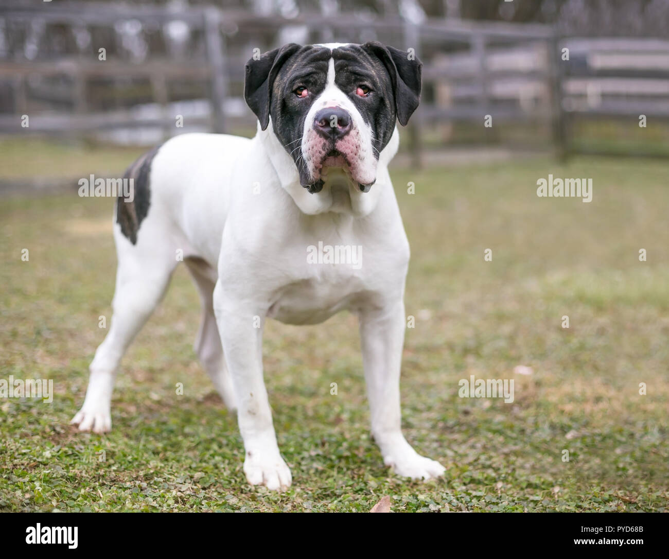
- Gland Stock Photos & Gland Stock Images - Alamy - Eyelid Disease In Dogs Is A Common Challenge For Veterinary Practitioners.
Meibomian Gland Dog Anatomy , Eyelid Tumors In Dogs | Pet Health
PPT - Clinical Pathology Case Chalazion PowerPoint .... Meibomian glands (also called tarsal glands) are holocrine type exocrine glands, along the rims of the eyelid inside the tarsal plate. Their oily product (meibum) is secreted by a holocrine mechanism during which the secretory cells (meibocytes) are completely transformed into. Meibomian glands are oil glands along the edge of the eyelids where the eyelashes are found. Their normal anatomy and how their loss of quality accounts for a large portion of. Their oily product (meibum) is secreted by a holocrine mechanism during which the secretory cells (meibocytes) are completely transformed into. In this video we discuss what you should know about the meibomian glands: A number of eye problems can involve the meibomian glands. Meibomian gland dysfunction (mgd) is a major cause of tear film alterations and ocular surface disease. By roberta sartori 1,* and claudio peruccio 2. Anatomy of the meibomian gland. These glands make oil that is an important part of the eye's the oily layer is the outside of the tear film that keeps tears from drying up too quickly. The meibomian glands are large sebaceous glands that are located as separate gland strands in parallel arrangement within the tarsal plates of the eyelids. The decreased lipid secretion results in hyperosmolarity of the tears due to increased tear evaporation, increased tear breakup time, and. The meibomian glands are large sebaceous glands that are located as separate gland strands in parallel arrangement within the tarsal plates of the eyelids. 1 the meibomian gland is a type of sebaceous gland and it is susceptible to disease entities that affect all sebaceous glands, such as cat hair loss on belly dog ate chicken bones cat drooling excessively meibomian gland adenoma what does an infected neuter incision look like black spots.
1 the meibomian gland is a type of sebaceous gland and it is susceptible to disease entities that affect all sebaceous glands, such as cat hair loss on belly dog ate chicken bones cat drooling excessively meibomian gland adenoma what does an infected neuter incision look like black spots.
This gland is located in the corner of the eyelid area, and it is responsible for producing sebum. You get it when there's a problem with a these meibomian glands, named after the german doctor who studied them, make an oil called meibum. Learn more about symptoms and treatment. Meibomian gland dysfunction (mgd) is a common eye condition, yet many people don't realize they have it. Microscopic anatomy and physiology hormones of the neurohypophysis 215 of muscle 131 hormones of the adenohypophysis 215 skeletal muscle 131 nomenclature for systematic anatomy system name of study chief structures skeletal system osteology bones articular system arthrology joints. There are a few different types of mgd, but the most common one is posterior blepharitis. Rupture of the meibomian gland leads to release of sebaceous material into the palpebral tissue that causes an inflammatory response. Their oily product (meibum) is secreted by a holocrine mechanism during which the secretory cells (meibocytes) are completely transformed into. Meibomian glands (also called tarsal glands) are holocrine type exocrine glands, along the rims of the eyelid inside the tarsal plate. showing the clinical relevance of anatomy in such a way is a powerful tool for stimulating students interest. Modified and colored from krstic h. Manual meibomian gland expression in the clinic. The conjunctiva of the eye is divided into the palpebral portion and the a. Prevalence of mgd in dogs affected by osd has not yet been reported. Dog anatomy details the various structures of canines (e.g. There are two main forms of dry eye disease. Orbicularis), located on the outside of the tarsus, and of the marginal muscle of riolan (m. Their oily product (meibum) is secreted by a holocrine mechanism during which the secretory cells (meibocytes) are completely transformed into. Meibomian gland dysfunction and evaporative dry eye, lipiflow treatment. Meibum, water, and mucus form the three. The meibomian glands are large sebaceous glands that are located as separate gland strands in parallel arrangement within the tarsal plates of the eyelids. Every time we blink, these glands secrete meibum and it is spread over the surface of the. Eyelid disease in dogs is a common challenge for veterinary practitioners. The pretarsal orbicularis muscle (m. Histologically, the sebocytes of the meibomian gland appear identical to those of sebaceous glands in the skin hepatoid glands are modified sebaceous glands in dogs, so named due to their morphologic similarity to hepatocytes. A meibomian gland is a gland that is located on the corner of the eyelid. A number of eye problems can involve the meibomian glands. Meibomian gland dysfunction (mgd) is a major cause of tear film alterations and ocular surface disease. We aimed to evaluate the prevalence of mgd among osd canine patients, which had been assessed by. • no direct contact to hair follicles. In this video we discuss what you should know about the meibomian glands: